
This is a detailed introduction to the Image Enhancer (I.I) for X-ray machines, which includes technical analysis, application scenarios, and industry trends. It is suitable as a medical device description or popular science article
1、 Definition and core functions of image intensifier (I.I)
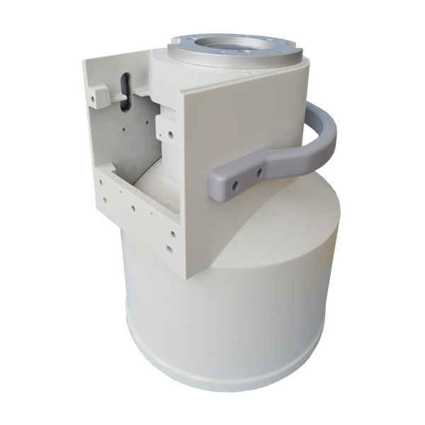
An image intensifier is the core component of X-ray machines, especially fluoroscopy equipment, used to convert invisible X-ray images into visible light images and significantly enhance image brightness, allowing doctors to observe dynamic images in real-time under low-dose radiation. Its core functions include:
-Brightness enhancement: Amplify weak X-ray signals thousands of times, reducing exposure requirements.
-Real time imaging: supports dynamic observation (such as cardiovascular intervention, gastrointestinal imaging).
-Radiation protection: Reduce the X-ray dose accepted by both doctors and patients.
2、 Structure and Working Principle
1. Core structure
-Input fluorescent screen: Convert X-rays into visible light (commonly using cesium iodide CsI material).
-Photocathode: converts visible light into electron flow.
-Electronic lens: accelerates and focuses electrons through a high-voltage electric field.
-Output fluorescent screen: Electronic impact produces high brightness visible light images.
-CCD/CMOS camera: converts optical signals into video signals (digital models).
2. Workflow
`X-ray → Input fluorescent screen (visible light) → Photocathode (electron) → Electron acceleration focusing → Output fluorescent screen (enhanced light signal) → Camera acquisition → Display`
3、 The core advantages of image intensifiers*
1. High sensitivity and low dose*
-Traditional I. I can reduce radiation dose to 1/10 of regular X-rays, making it suitable for long-term fluoroscopy (such as surgical navigation).
2. Real time dynamic imaging
-Supports frame rates above 30fps to capture dynamic processes such as organ movement and contrast agent flow.
3. Cost effectiveness
-Compared to digital flat panel detectors (FPDs), the procurement cost of I.I systems is 40% -60% lower.
4、 Main technical parameters and performance indicators
1. Input screen size
-Common specifications: 6 inches, 9 inches, 12 inches (the larger the field of view, the wider the field of view, but the resolution may decrease).
2. Brightness gain
-Typical value: 5000-10000 cd/m ², determining imaging clarity at low doses.
3. Resolution
-Center resolution ≥ 4 lp/mm (line pairs/mm), edge resolution ≥ 3 lp/mm.
4. Quantum Detection Efficiency (DQE)
-The DQE of high-quality I. I can reach over 65%, reducing image noise.
5、 Maintenance and common problems
1. Daily maintenance
-Regularly calibrate geometric distortion (once a month), clean the input screen (using anhydrous ethanol).
2. Typical faults
-Edge blurring: The voltage of the electronic lens is offset and needs to be recalibrated.
-Brightness decrease: The input screen is aging and the fluorescent layer needs to be replaced.
3. Life extension techniques
-Avoid prolonged high-dose exposure and turn off the high-voltage power when shutting down.
Despite the rapid development of digital flat panel detectors, image intensifiers are still irreplaceable in real-time dynamic imaging and low-cost fields. Medical institutions should choose equipment based on surgical type, budget, and radiation management needs, while industrial users can prioritize the cost-effectiveness advantage of I. I.
Newheek was founded in 1999 and has been focusing on the X-ray field for more than 20 years. It has a complete range of X-ray machines with various specifications and a complete production line of image intensifiers. If you are interested in watching movies, please contact me, whatsapp+86 19953639012
Post time: Apr-26-2025